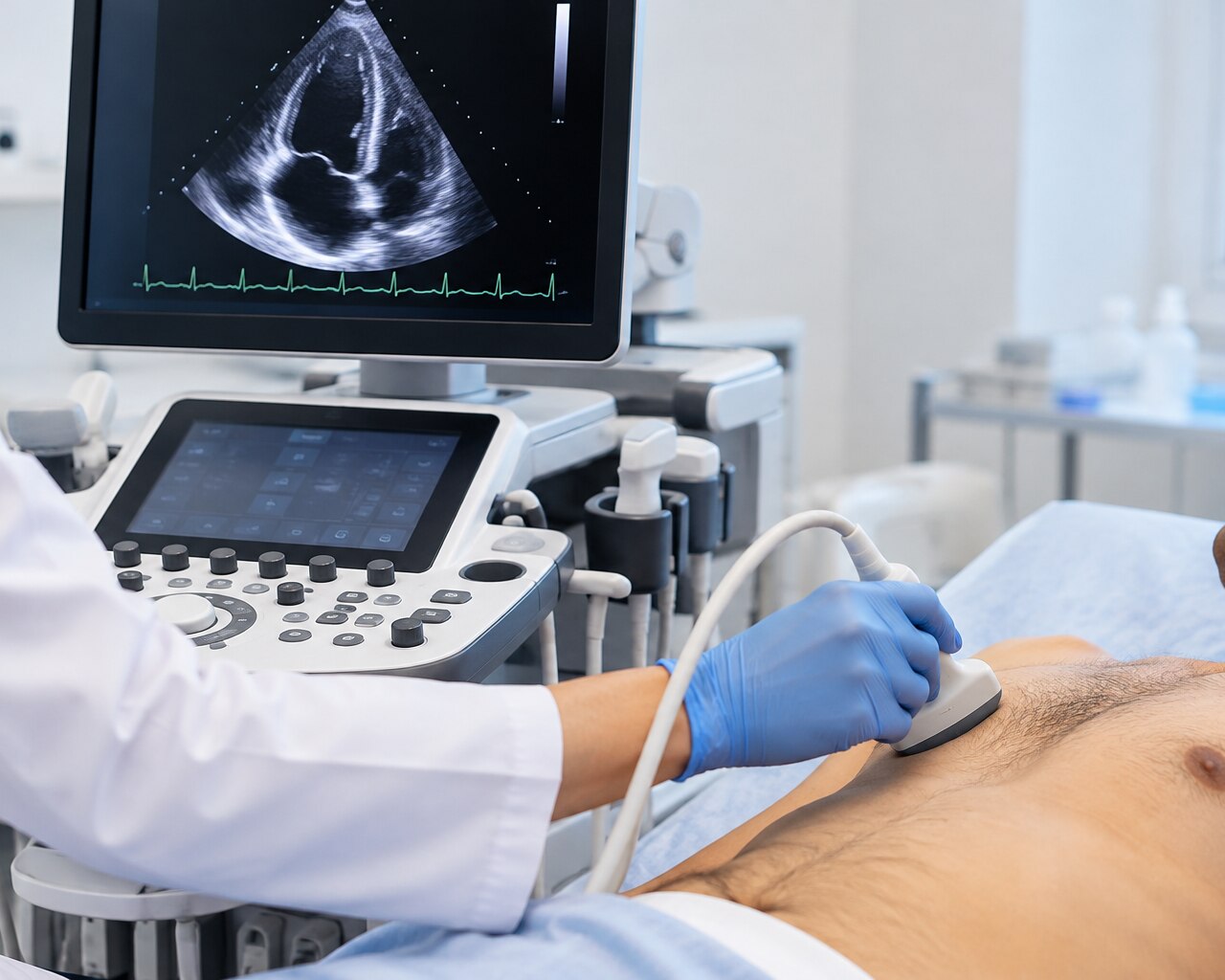
Breathlessness, chest discomfort, swelling, fatigue, a murmur or an abnormal ECG do not all point to the same heart test. By matching your symptom, suspected condition and the question your clinician needs answered, you can understand whether you need a resting echo, stress echo, transoesophageal echo or another form of imaging.
Key takeaways
- A resting TTE is usually the first echo for breathlessness, swelling, murmurs, or abnormal ECGs.
- A stress echo assesses heart function and blood flow during exercise or medicine-induced stress.
- TEE, contrast, and 3D echo answer detailed questions that standard TTE cannot.
- Confirm the test type, preparation, report timing, and cardiologist review before booking.
Which heart symptoms usually call for a resting echocardiogram?
A resting transthoracic echocardiogram (TTE), also called a 2D echo, is usually the starting test for breathlessness, leg swelling, fatigue, a heart murmur, chest discomfort, suspected heart failure, a previous heart attack, suspected valve disease or an abnormal ECG. It uses sound waves, not X-rays.
A sonographer moves a probe over your chest to assess chamber size, pumping function, valve movement, wall motion, blood flow, pressure estimates and selected structural abnormalities. Doppler and colour-flow imaging show the direction and speed of blood, not merely a picture of the heart.
| Symptom or finding | What a resting TTE can assess | What the result may help investigate |
|---|---|---|
| Breathlessness, fatigue or leg swelling | Pumping function, chamber size and pressure estimates | Heart failure or raised pressures |
| Murmur or suspected valve disease | Valve movement and blood-flow patterns | Narrowing or leakage |
| Chest discomfort or previous heart attack | Wall motion and pumping function | Damage or reduced movement |
| Abnormal ECG | Structure and mechanical function | A finding needing further assessment |
A standard TTE usually requires no fasting, injection or recovery period. If you are asking, “which echocardiography test do i need,” the decision belongs with a clinician; a normal or technically limited scan cannot exclude every rhythm disorder, lung problem or early coronary disease.
Seek emergency assessment for sudden chest pressure, severe breathlessness, fainting, new weakness or confusion. Do not wait for a routine echo appointment.
When is a stress echocardiogram more useful than a resting echo?
Exertional chest pain or suspected reduced blood flow makes a stress echocardiogram more useful than a resting echo because it tests the heart under demand. A resting scan can show normal pumping at rest while missing a blood-supply problem that appears only during exertion. Symptoms alone do not determine the correct test.
An exercise stress echo takes images at rest and immediately after walking on a treadmill or cycling. If you cannot exercise adequately, a medication-induced study commonly uses dobutamine to increase the heart’s workload. New regional wall-motion changes during stress can indicate inadequate blood supply, but the test does not directly show coronary narrowing.
Selection depends on your pretest likelihood of coronary disease, resting ECG, earlier test results, mobility and the local team’s expertise. Seek medical review before exercise testing if you have:
- Unstable or worsening chest symptoms
- Severe illness
- A major mobility limitation
- An inability to exercise enough for the protocol
Stress echo assesses heart function under demand; the alternatives answer different questions.
| Test | Main information | Key limitation |
|---|---|---|
| Stress echo | Wall motion and pumping during stress | Does not map coronary anatomy directly |
| ECG or treadmill test | Electrical activity, symptoms and exercise response | Gives less direct information about heart structure and wall motion |
| CT coronary angiogram | Coronary-artery anatomy and narrowing | Does not assess valve motion in the same way |
| Invasive angiography | Detailed coronary anatomy during catheter testing | Invasive and not a routine first choice for every symptom |
When does a transoesophageal, contrast or 3D echo answer a question that TTE cannot?
Choose the advanced echo according to the question your clinician needs answered, not because one scan is automatically more complete than another.
| Option | What it shows best | When it applies |
|---|---|---|
| Transoesophageal echo (TEE) | Close views of the left atrial appendage, selected valves and aorta | Suspected clot, infective endocarditis, prosthetic-valve malfunction or a procedural complication |
| Contrast echo | Sharper heart-wall borders and pumping-function measurements | Body habitus, lung interference or difficult image windows make a standard TTE unclear |
| Three-dimensional echo | Valve shape and chamber-volume measurements | Detailed valve planning or more precise volume assessment when image quality supports it |
TEE places a flexible probe through your mouth into the oesophagus, behind the heart. You must fast beforehand; the team numbs your throat and commonly gives sedation, so arrange an escort and do not drive afterward. Tell them about swallowing difficulty or oesophageal disease.
Throat discomfort is common, while aspiration or injury to the teeth, throat or oesophagus is uncommon but possible.
Contrast echo improves endocardial-border definition and helps estimate pumping function when ordinary images are inadequate. It does not replace TEE for the left atrial appendage, valves or aorta, and it does not show coronary-artery narrowing.
Three-dimensional echo is a selected tool, not a requirement for every routine scan. A technically limited result can still leave rhythm, lung or early coronary problems unanswered, requiring another test.
How do other heart tests compare with echocardiography?
The right test depends on the question: electrical activity, exercise response, coronary-artery narrowing, tissue damage or heart structure.
| Test | What it shows | When it fits |
|---|---|---|
| Echocardiogram | Valve shape and motion, chamber size, pumping and blood flow | Suspected structural or blood-flow problem |
| ECG | Electrical activity, rhythm and conduction | Palpitations, fainting or an abnormal pulse; it does not show valve shape or chamber pumping |
| Treadmill stress test | ECG and symptoms during exertion | Assessing exercise response when walking is safe; it gives less direct structural information |
| CT coronary angiography | Coronary-artery narrowing using X-rays and contrast | Suspected coronary disease; it does not measure valve motion like an echo |
| Cardiac MRI | Heart volumes, tissue, scar and inflammation | Complex muscle disease or unclear findings; the scan takes longer and requires suitability checks |
A stress echocardiogram combines exercise or dobutamine with ultrasound images, looking for stress-induced changes in heart-wall movement. It is not automatically the best test for chest pain; your pretest likelihood, resting ECG, exercise ability, earlier results and local expertise guide the choice.
Fetal echocardiography is a targeted examination of an unborn baby’s heart when screening or family risk raises concern. It is separate from a routine obstetric ultrasound.
A normal echo does not explain every symptom. Palpitations, persistent breathlessness or chest pain may require rhythm monitoring, blood tests, chest imaging, coronary imaging or specialist assessment.
What should you confirm before booking an echo in Dombivli?
Do not book from a service list alone: confirm which echocardiogram answers your clinician’s question. Ask whether you need TTE, stress echo, TEE, contrast echo, 3D echo, or fetal or pediatric echo, and whether a cardiologist will interpret the images.
| Option | What it involves | Confirm before booking |
|---|---|---|
| TTE | Probe over the chest at rest | Usually no fasting, injection or recovery |
| Stress echo | Images during exercise or after dobutamine | Exercise ability, medication instructions and monitoring |
| TEE | Probe passed into the oesophagus | Fasting, sedation, escort and swallowing or oesophageal problems |
| Contrast echo | Injected contrast improves unclear heart borders | Injection suitability and observation arrangements |
| 3D echo | Three-dimensional views of selected structures | Whether it changes the clinical answer |
| Fetal or pediatric echo | Targeted imaging for an unborn baby, infant or child | Specialist availability and age-specific reporting |
Before travelling, ask the diagnostic department:
- What preparation applies, including fasting, medication changes, sedation and an escort?
- Will the facility provide exercise testing, continuous monitoring and emergency support for a stress echo?
- Who reports the images, when will the report be ready, and can your referring clinician receive it?
The ordering clinician should choose the test after reviewing your symptoms, examination, ECG, medical history and ability to exercise.
A cardiology consultation in Dombivli East at Aurindam Hospital can connect those findings with the appropriate examination; its diagnostic department can confirm preparation and availability for echocardiography options Dombivli, heart ultrasound near Dombivli or an echo test hospital Dombivli search.
For sudden chest pressure, severe breathlessness, fainting, new weakness or confusion, contact emergency services instead of booking outpatient imaging.
Related services
Cardiology & Echocardiography Cardiology is the branch of medicine that deals with the diagnosis, treatment, and prevention of diseases related to the heart and blood vessels... View service → |
Stress Test/Stress Echo A stress echo test, or stress echocardiogram, is a non-invasive procedure used to evaluate how well your heart functions under stress, either through... View service → |
Frequently asked questions
Which heart symptoms usually call for a resting echocardiogram?
A resting transthoracic echocardiogram is commonly used for breathlessness, leg swelling, fatigue, a heart murmur, chest discomfort, suspected heart failure, previous heart attack, suspected valve disease, or an abnormal ECG.
When is a stress echocardiogram more useful than a resting echo?
A stress echocardiogram compares heart function at rest and during exercise or medication-induced stress, helping assess symptoms linked to exertion and possible reduced blood flow.
When does a transoesophageal, contrast or 3D echo answer a question that TTE cannot?
Transoesophageal echo provides closer views through the oesophagus, contrast improves poorly seen chambers or borders, and 3D echo adds detailed views for selected valve and structural assessments.
How do other heart tests compare with echocardiography?
An ECG records electrical activity, a stress ECG monitors electrical changes during exertion, cardiac CT shows anatomy with X-rays, cardiac MRI provides detailed tissue and structure images, and blood tests measure markers such as troponin. Echocardiography shows moving heart chambers, valves, blood flow, and pumping function without X-rays.
What should you confirm before booking an echo in Dombivli?
Confirm whether you need a resting TTE, stress echo, TEE, contrast echo, or 3D study; ask about preparation, report timing, cardiologist review, and whether the facility handles urgent symptoms.
Keywords